Perettiite-(Y)
A new mineral from Momeik, Myanmar
by Danisi, Rosa Micaela; Armbruster, Thomas; Libowitzky, Eugen; Wang, Hao A.O.; Günther, Detlef; Nagashima, Mariko; Reusser, Eric; Bieri, Willy
European Journal of Mineralogy Volume 27 Number 6 (2015), p. 793 - 803, published: Dec 14, 2015
Keywords: Perettiite-(Y); Y³⁺₂ Mn²⁺₄ Fe²⁺ [ Si₂ B₈ O₂₄ ]; crystal structure; new mineral; borosilicate; Momeik; Myanmar.
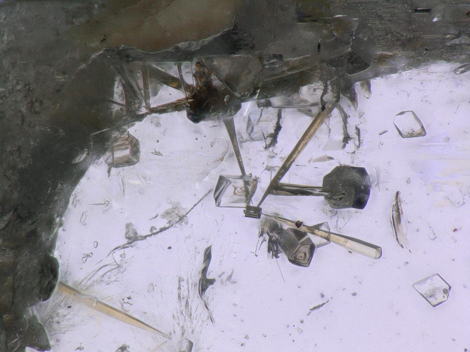
The new mineral perettiite-(Y), (IMA 2014-109), ideally Y3+2Mn2+4Fe2+[Si2B8O24] as been discovered in the region of Momeik, north of Mogok, Myanmar. It was found as inclusions in perfect gemmy colorless transparent phenakite crystals originated from isolated pegmatite pockets of granitic pegmatites intruding large peridotite body. Of the stock of ~10 000 phenakite centimeter-sized crystals, only 15 were containing inclusions of perettiite-(Y). Other inclusions in phenakite are schorl, tusionite, columbite-(Mn), albite, fluorapatite, and lazulite.
Phenakite crystals found in pockets with quartz, feldspar, and schorl. Neighborhood pegmatites contain famous mushroom and botryoidal tourmalines, hambergite, petalite, beryl (aquamarine and morganite), pollucite, danburite, topaz, almandine-spessartine, biotite, magnetite, lepidolite, hubnerite-ferberite, and cassiterite. Perettiite-(Y) forms yellow needles elongated by [010] up to a few millimeters long and up to 0.2 mm thick. Observed crystal forms are {100} and {001}. Crystals are intimately twinned. The mineral has white streak and vitreous luster. It is brittle, with irregular fracture and good {010} cleavage. The micro-indentation hardness is VHN300 = 100 (100–110) kg/mm2 corresponding to ~7 of Mohs scale. Density was not measured due to intergrowth with phenakite; Dcalc = 4.533 g/cm3. Perettiite-(Y) is optically biaxial, α = 1.82(1), γ = 1.84(1) (589 nm).
Due to intimate twinning, the crystal appears conoscopically uniaxial with diffuse isogyre cross, thus the optical character 2V and β could not be estimated. Under crossed polars the mineral shows on (010) sections a characteristic hourglass pattern (similar to apophyllite) with undulatory extinction. Single crystal Raman spectra (488 nm Ar-ion laser) exhibit multiple and intense luminescence emission lines (1200–1600 and 1800–2700 cm−1) probably related to lanthanoids content. A long-term exposure allows to identify vibrations at ~1000, 700–800, and <500 cm−1 assigned to common borosilicate stretching, bending and lattice modes. The absence of bands at 3000–3700 cm−1 do not confirm the presence of H2O/OH. Single-crystal FTIR spectra exhibit an intense O–H stretching band at 2750–3750 cm−1.
Quantitative calculation yields a maximum hydroxyl/water content equivalent to 0.1 wt% of H2O. The chemical data by LA-ICP-MS [wt% (range)] are followed (where determined) by electron probe WDS data (bolded) for 2 samples: Li2O 0.32 (0.24–0.38); BeO 0.75 (0.66–0.82); B2O3 24.86 (24.61–25.12); MgO 0.27 (0.23–0.29) 0.56, 0.44; Al2O3 0.56 (0.48–0.62); SiO2 11.26 (10.42–12.02), 11.88, 11.94; CaO 2.02 (1.82–2.27), 1.66, 2.00; MnO 22.06 (21.04–23.56), 22.95, 21.20; FeO 4.89 (4.62–5.15), 4.62, 4.52; Y2O3 22.32 (21.81–23.04), 19.00, 20.99; ZrO2 0.19 (0.17–0.20); Sm2O3 0.24 (0.23–0.27); Gd2O3 0.71 (0.66–0.80), n.d., 1.42; Tb2O3 0.29 (0.28–0.31); DY2O3 2.62 (2.45–2.75), n.d., 2.14; Ho2O3 0.53 (0.50–0.55); Er2O3 1.78 (1.73–1.92), n.d., 1.71; Tm2O3 0.33 (0.32–0.37); Yb2O3 2.85 (2.59–3.24), n.d., 2.68; Lu2O3 0.38 (0.35–0.42); ThO2 0.33 (0.30–0.41); total 99.56.
The empirical formula based on 24 O pfu is Y2.06Ln0.53Zr0.02Th0.01Mn3.24Ca0.38Fe0.71Mg0.07Al0.11Li0.22Si1.95B7.44Be0.31O24.
The strongest lines in the X-ray powder pattern [d Å (I%; hkl)] are 4.63 (52; 010), 4.08 (28; 301,103), 3.74 (20; 210), 3.05 (100; 113,311,303), 2.64 (67; 410,014), 2.54 (60; 313), 2.12 (23; 600,006), 1.87 (33; 420,024), 1.84 (52; 415,323), 1.57 (20; 026,620), 1.44 (25; 133,331).
The single-crystal X-ray study shows a tetragonal X-ray diffraction pattern, but the structure could only be solved as a 50/50 pseudo-merohedral orthorhombic twin with the a = c. The structure was solved by direct methods and refined to R1 = 0.017 on the basis of 1814 unique I > 2σ(I) reflections in space group Pmna with a = 12.8252(5), b = 4.6187(2), c = 12.8252(5) Å, V = 759.71 Å3, Z = 2. The crystal structure of perettiite-(Y) has two eightfold-coordinated sites: one dominated by Y and Ln and the other by Mn2+ (with additional Ca2+ and Y3+). An octahedral site is occupied by (Fe2+, Mg) with additional Li+. These cation sites form an interlayer between two borosilicate tetrahedral Si2B8O24 sheets parallel to (010) formed by 4-, 5- and 8-membered rings. B shows minor replacement by Be. The structural relationships with other species with similar tetrahedral sheets are discussed.
The mineral was named after the mineralogist and gemologist Adolf Peretti (b. 1957), mineralogist and head of GRS Gemresearch Swisslab, who first recognized inclusions in phenakite. The holotype specimen is deposited in the Museum of Natural History Bern, Switzerland.
© 2024, GRS GemResearch Swisslab AG